Deafness and hearing loss toolkit
Guidance for GPs on the care of patients dealing with deafness and hearing loss.
Interpretation of Investigations
Please note an audiologist/AVM/ENT will send a summary/copy of the basic investigations below to you after the assessment has taken place.
Otoscopy
The British Society of Audiology produced a detailed procedure for ear examination.
Tuning Forks
Provides preliminary diagnostic information.
Rinne
- Comparison of air conduction (AC) and bone conduction (BC) sensitivity
- Tuning fork is alternated between entrance of ear canal and mastoid process
- Rinne Positive: Tuning fork is louder via AC= sensorineural hearing loss (SNHL)
- Rinne Negative: Tuning fork is louder on mastoid= conductive hearing loss (CHL)
Weber
- Test of lateralisation and therefore may be used for patients who report unilateral hearing loss.
- The tuning fork is placed midline on the patients’ forehead.
- If sound lateralises to ear with loss= CHL as the improved BC is due to the occlusion effect.
- If soundlateralises to ear without loss=SNHL or mixed hearing loss (MHL) as the best cochlea is detecting the signal. In normal hearing= midline
Types of hearing loss
- CHL is a result of dysfunction in the middle or outer ear (BC >AC)
- SNHL is a result of cochlea damage (sensory) and/or neural (8th nerve) (BC = AC)
- Mixed hearing loss is a combination of dysfunction in middle/outer ear and cochlea/8th nerve (A certain amount of AC and BC loss)
- Central hearing loss refers to everything in the auditory cortex (Brain) whereas peripheral hearing loss is the result of everything before the brain (outer, middle, inner ear)
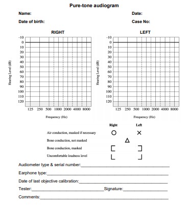
Interpreting a Pure-tone Audiogram (PTA)
- An audiogram is a plot of frequency in Hertz (Hz) against intensity measured in decibels hearing level (dB HL). The frequency range for a PTA is 250 Hz to 8000Hz as this range of frequencies is similar to the range important for speech understanding.
- The intensity ranges from -10dB HL to 120dB HL. Thresholds (lowest acoustic intensity perceived at a given frequency) for both the right and left ear are plotted based on the frequency. Refer to image below.
- Masked AC/BC thresholds are an accurate assessment of the test ear.
- Further interpretation and explanations on Pure-tone Audiogram are available on the BSA website:
| Descriptor | Average hearing threshold levels (dB HL) |
|---|---|
| Normal Hearing | < 20 |
| Mild hearing loss | 21 - 40 |
| Moderate hearing loss | 41 - 70 |
| Severe hearing loss | 71 - 95 |
| Profound hearing loss | > 95 |
Interpreting a Tympanogram
- Tympanogram is graphic display of tympanic membrane compliance (ml or cm3) as a function of pressure changes in the external auditory meatus. See figure 1.
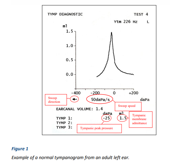
- Tympanometry is sensitive to middle ear effusion, cholesteatoma, ossicular adhesions, and space occupying lesions in contact with the eardrum, ossicular discontinuity, perforations and ear canal occlusions.
- The shape of the tympanogram should also be described and simple descriptions such as ‘normal’, ‘rounded’, ‘flat’, ‘wide’ or ‘W-shaped’.
- The BSA has further interpretation and explanations on Tympanometry.
Examples of Types of Hearing Loss
Conductive Hearing Loss
- An audiogram illustrating a mild conductive hearing loss in the right ear. The air-conduction thresholds for the right ear are shown as O’s and the masked, bone-conduction thresholds are shown as brackets.
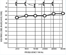
- Examples of a conductive hearing loss included a perforated tympanic membrane, and otitis media with effusion etc.
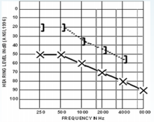
Sensorineural Hearing Loss
- An audiogram illustrating a mild-moderate, high-frequency sensorineural hearing loss in the right ear.
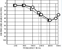
- Examples of a SNHL includes presbycusis, an acoustic neuroma, Meniere’s disease, and noise-induced hearing loss etc.
Mixed hearing loss
- An audiogram illustrating a moderate-to-severe, mixed hearing loss in the left ear. Both the air-conduction thresholds (X’s) and the bone-conduction thresholds (brackets) indicate hearing loss.
- Examples of mixed hearing loss includes otosclerosis, a SNHL with a perforation/otitis media with effusion.